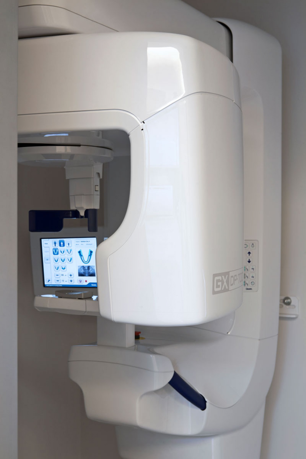
2D and 3D X-ray
The use of X-ray technology in dentistry is fundamental for a correct diagnosis. Our clinic is provided with state-of-the-art last-generation X-ray equipment, undergoing periodical maintenance and quality control to test conformity and radiation exposition.
These are the diagnostic examinations offered in our clinic:
INTRA-ORAL X-RAYS: 1 or more X-ray images of single teeth showing structure, roots and periodontal support, taken from different points of view.
When a Full-Mouth X-ray (FMX) is taken, all intra-oral images are displayed on a specific instrument allowing the clinician to have a complete view on the patient’s situation.
By analyzing the X-rays and the parameters collected during the patient’s examination, the clinician is able to present a comprehensive treatment plan right on the first appointment.
Bite-wing X-Rays are intra-oral pictures covering up to 8 teeth and allowing to simultaneously evaluate the coronal area (the exposed part of the tooth) and the adequacy of the periodontal support. If regularly taken on clinical appointments, these X-rays provide the clinician with a follow-up of the health state of the patient’s teeth, fillings and prosthesis.
CBCT (Cone Beam Computer Tomography): usually referred to as CT scan, the Cone Beam X-ray is a 3D examination of anatomical structures and its possible lesions. Our clinic is provided with a state-of-the-art CT scan providing excellent 3D X-rays while reducing the exposure to radiation.
The use of a CBCT is essential to evaluate the health state and adequacy of bone support before an implant surgery, as well as to determine the exact position of the wisdom teeth before extraction.
ORTHOPANTOMOGRAM: also known as “Panoramic X-ray”, this exam provides an overall view of the maxillary bones and is useful - together with teleradiographies - to elaborate treatment plans for patients undergoing orthodontics.
TELERADIOGRAPHY: this X-ray image provides an overall view of the anatomy of the skull: the parameters measured are useful to evaluate the reciprocal position of the arches and detect possible misplacements of the occlusal plane, which will have to be corrected by undergoing an orthodontic or orthognatic treatment.FF